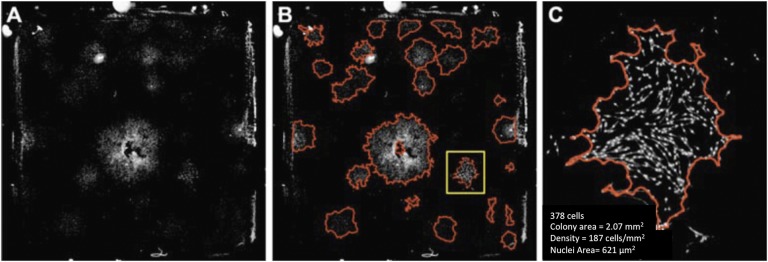
Fig. 2.
Fig. 2-A Large field-of-view images (480 image tiles stitched together) of CTPs seeded on 20.5 × 20.5-mm chamber slides and stained with DAPI. The images shown here have been background-corrected (flattening illumination for each tile) and processed for the removal of artifacts. Fig. 2-B Images processed for colony segmentation (red outline) using an automated algorithm. Fig. 2-C Magnified colony (delineated by yellow in Fig. 2-B) indicating quantitative parameters that may be extracted. (Reprinted, with permission, from ASTM F2944-12 Standard Test Method for Automated Colony Forming Unit (CFU) Assays—Image Acquisition and Analysis Method for Enumerating and Characterizing Cells and Colonies in Culture, copyright ASTM International, 100 Barr Harbor Drive, West Conshohocken, PA 19428. A copy of the complete standard may be obtained from ASTM, www.astm.org.)

