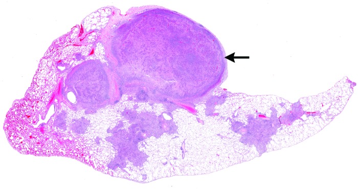
Figure 3.

Histologic section of macaque lung, demonstrating multifocal to coalescing areas of consolidation throughout the section. Note the severely affected nodular area (top) that is protrusively distorting the pleura. Hematoxylin and eosin stain; magnification, 3×.
