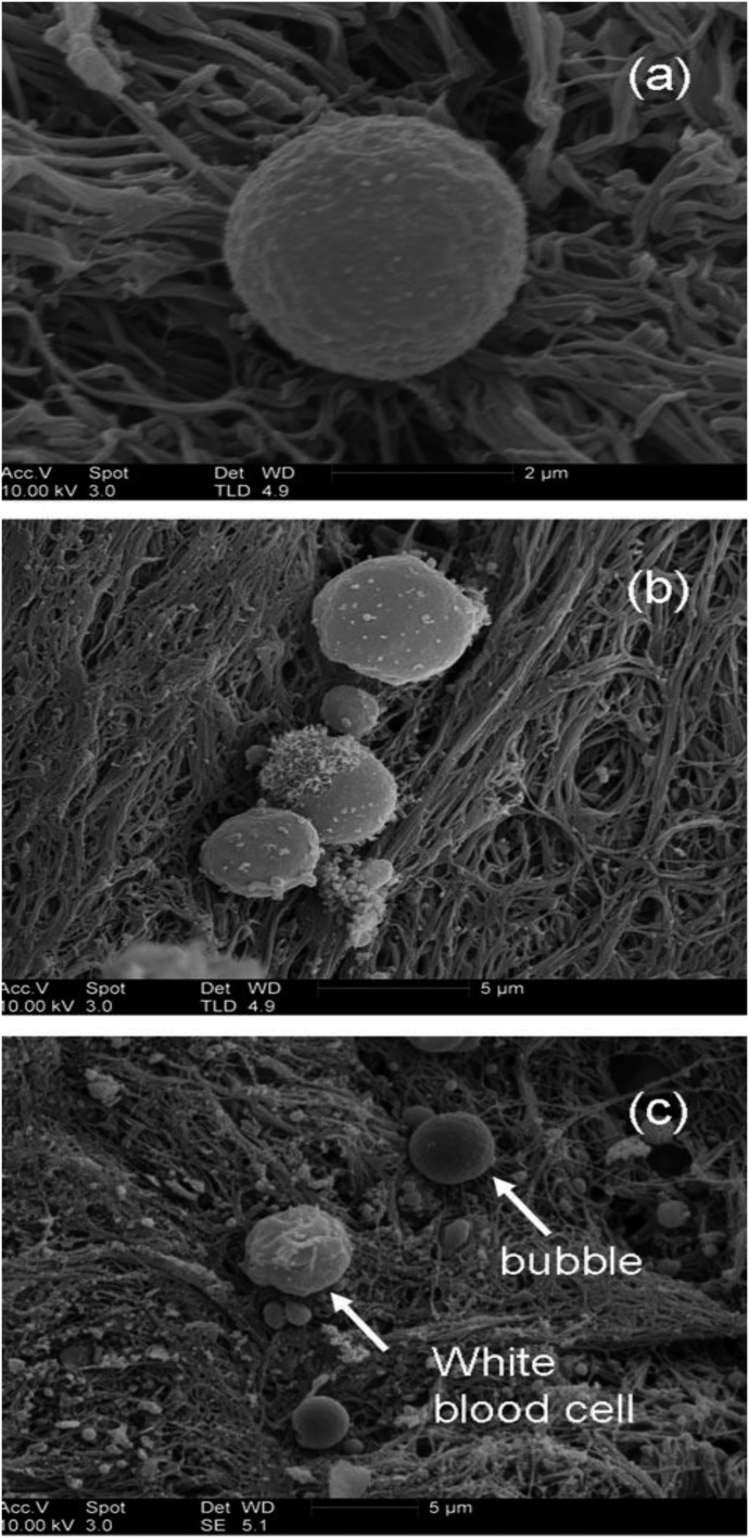
Figure 3.
SEM images obtained from thrombus exposed to Abciximab microbubbles. (A) A single bubble. (B) Three bubbles. The largest bubble is 4 μ in diameter, which is too big to be a platelet and does not resemble any other blood cell. (C) A smooth, spherical bubble, and a white blood cell. (Martin MJ, Chung EML, et al. Enhanced detection of thromboemboli with the use of targeted microbubbles. Stroke 2007;38:2726–32).

