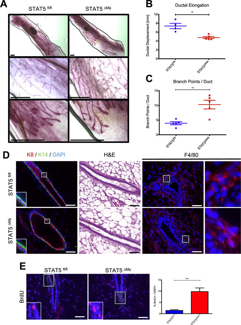
Fig. 2.

STAT5 deletion in macrophages disrupts normal mammary gland development. A) [Top] Whole mount analysis of mammary glands. [Middle] High-power magnification of mammary ducts. [Bottom] High-power magnification of terminal end buds. B) Quantification of ductal elongation in mouse mammary glands. C) Analysis of epithelial branchpoints per mammary duct. Scale bars represent 1 mm. D) [Left] Paraffin-embedded mammary gland sections were stained for K8 (red), K14 (green), and DAPI (blue). Regions identified in squares magnified in insets. [Middle] H & E staining of mammary gland sections. [Right] Paraffin-embedded mammary gland sections were stained for F4/80 (red) and DAPI (blue). Regions identified in squares magnified in insets. E) [Left] Paraffin-embedded mammary gland sections were stained for BrdU (red) to assess proliferation and counterstained with DAPI (blue). Regions identified in squares magnified in insets. [Right] Quantification of proliferating cells normalized to total number of DAPI+ cells. Scale bars represent 50 μm. n=4 (STAT5fl/fl) and n=5 (STAT5ΔMϕ), **p < 0.01, ****p < 0.0001.
