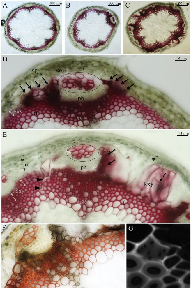
Fig 3. Inflorescence cross sections at intermediate and mature stages of development.
Cross sections through the basal region of the infloresence stem for wild type (A) and mur1-1 (B,D) at an intermediate stage of development. Cross sections of the basal region of the stem of mur1-1 at a mature stage of development (C,E) stained with phloroglucinol-HCl or with the Maüle reagent (F). Phloem sclereids are encircled on D and E and abnormal lignified cells rich in aldehyde compounds are indicated by black arrows. The pitted cell wall of regenerative xylem and the fragmentation of the sclerenchyma cylinder are indicated by black arrow heads and dotted arrows respectively. The cortex cell wall layer are indicated by asterisks. Observation of the perforation plate of a regenerative xylem cell observed by confocal microscopy (G). Ep: epidermis, co: cortex, ph: phloem, xy: xylem, Sxy: secondary xylem, Rxy: regenerative xylem, if: interfascicular fibers.

