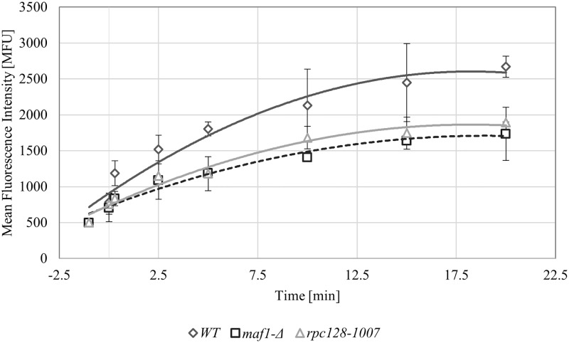
Fig 3. Glucose flux is present in all tested strains.
High-affinity glucose transport was measured in the WT (MB159-4D), rpc128-1007 and maf1-Δ strains. The assay was performed using fluorescently labeled glucose 2-NDBG. The cells were grown to exponential phase of A600 ≈ 1.0 in 2% glucose-rich medium (YPD), transferred to SC medium supplemented with amino acids without a carbon source and incubated for 10 min at 30°C. The uptake of 2-NDBG (1 mM concentration) was performed over time. As a control, cell suspensions without fluorescently labeled glucose were assessed. The results are expressed as the mean fluorescence intensity (MFU) of four independent biological replicates with standard deviations.

