
Figure 4.
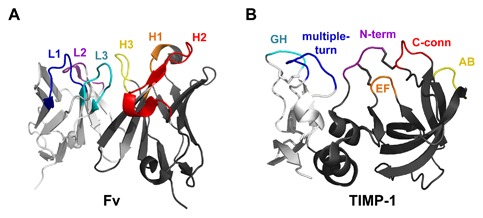
Similarities between antibody Fv and TIMP binding interfaces. (A) An Fv binds to an antigen through three loops of the heavy chain (dark gray): complementarity‐determining regions (CDRs) H1 (orange), H2 (red), and H3 (yellow), and three loops of the light chain (light gray): CDRs L1 (blue), L2 (purple), and L3 (cyan). Coordinates are from PDB ID: 1YQV [Cohen et al., 2005]. (B) TIMPs, like antibodies, recognize the MMP target using a broad interface comprised of multiple loops spread across two domains, including segments of the N‐terminal domain (dark gray): N‐terminus (purple), AB loop (yellow), C‐connector loop (red), and EF loop (orange), and segments of the C‐terminal domain (light gray): GH loop (cyan) and multiple‐turn loop (blue). Coordinates are from PDB ID: 1UEA [Gomis‐Ruth et al., 1997]. Figure was generated using PyMOL (Schrodinger, LLC).