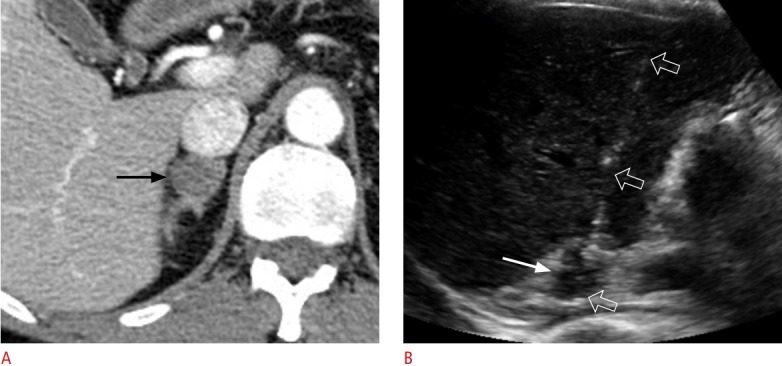
Fig. 7. A 59-year-old woman with schwannoma.

A. Contrast-enhanced axial computed tomography shows a 2.1-cm solid tumour (arrow) in the right adrenal gland. The lesion has a low percentage wash of contrast material on adrenal computed tomography. She was diagnosed with lung cancer. B. Transhepatic ultrasonography shows the lesion (arrow), which was targeted with an 18-gauge biopsy needle (open arrows). This histologic diagnosis was schwannoma.
