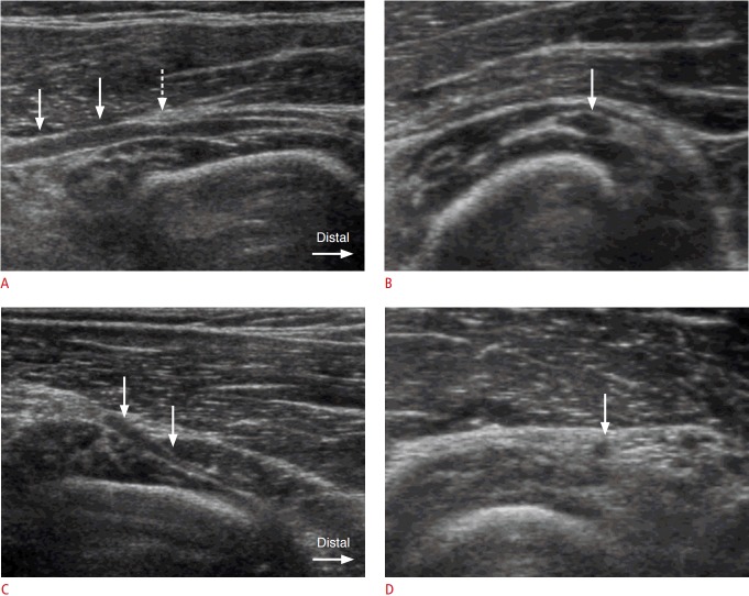
Fig. 1. A 42-year-old man with swelling of the posterior interosseous nerve (PIN) proximal to the supinator muscle.
A. Longitudinal ultrasonogrphy demonstrates hypoechoic swelling of the left PIN (arrows) proximal to the supinator muscle. Compression by the hyperechoic thickened arcade of Fröhse (dashed arrow) is noted. B. Axial ultrasonography shows swelling of the left PIN (arrow) at a level just proximal to the arcade of Fröhse. The thickness of the swollen PIN was measured as 2.4 mm in anteroposterior diameter. C, D. Ultrasonography of the contralateral asymptomatic right forearm shows a gradual tapering appearance of the PIN (arrows) in the longitudinal scan (C) with a normal thickness (1.0 mm in anteroposterior diameter) of the PIN (arrow) in an axial scan (D).

