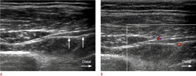
Fig. 3. A 49-year-old woman with swelling of the posterior interosseous nerve (PIN) distal to the supinator muscle.
A. Gray-scale ultrasonography shows hypoechoic swelling of the PIN (arrows) as it exits from the supinator canal (S). B. Color Doppler image demonstrates increased perineural vascularity at the site of hypoechoic swelling of the PIN.

