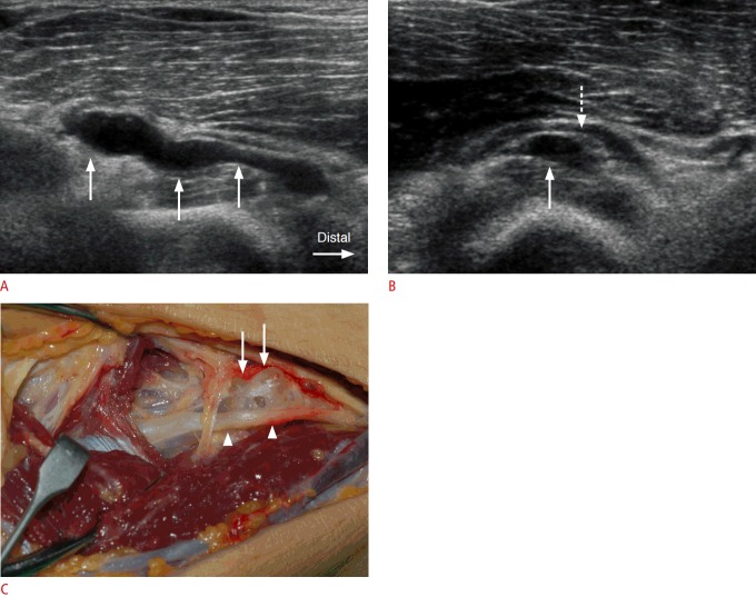
Fig. 4. A 54-year-old man with a ganglion cyst in the supinator canal.
A. A tubular, lobulated, anechoic ganglion cyst (arrows) is demonstrated from the site just proximal to the supinator muscle to the proximal supinator canal in a longitudinal ultrasonography. B. The posterior interosseous nerve (PIN) (dashed arrow) is displaced and followed due to an anechoic ganglion cyst (arrow) in the entrance site to the supinator canal in an axial ultrasonography. C. Surgical photograph demonstrates a ganglion cyst (arrows) and displaced PIN (arrowheads).

