
Fig. 1. Enhanced neck computed tomography findings.
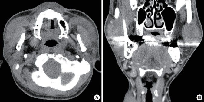
The masses were located between the masseter muscles and platysma, adjacent to the parotid gland, with nonhomogeneous mild enhancement and lymphadenopathy at both levels IA and IB. An axial view (A) and coronal view (B).