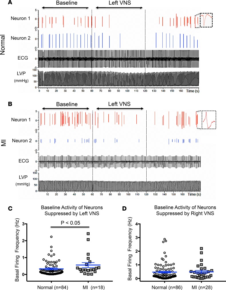
Figure 3. Recordings from cardiac neurons that are suppressed by VNS.
(A) Example of direct neuronal recordings from the VIV fat pad ganglia of a normal heart showing 2 neurons that decrease their firing activity with VNS and were therefore classified as postganglionic parasympathetic neurons. Each neuron was identified and classified by its unique neuronal waveform (inset). (B) Example of neuronal recordings from the VIV fat pad of an infarcted heart demonstrating 2 neurons that also decreased their firing frequency with VNS. Basal activity in the minute prior to VNS compared with during VNS was used to identify parasympathetic neurons. (C) The basal (prestimulation) activity of parasympathetic neurons that were suppressed by left VNS is reduced in MI compared with normal hearts (P < 0.05, unpaired Student’s t test, n = 84 neurons from 15 normal and n = 18 neurons from infarcted animals). (D) Basal activity of neurons that are suppressed by right VNS is unchanged in normal vs. infarcted hearts (P = 0.4, unpaired Student’s t test, n = 86 neurons from 15 normal and n = 28 neurons from 10 infarcted animals). LVP, left ventricular pressure; MI, myocardial infarction; VNS, vagal nerve stimulation.

