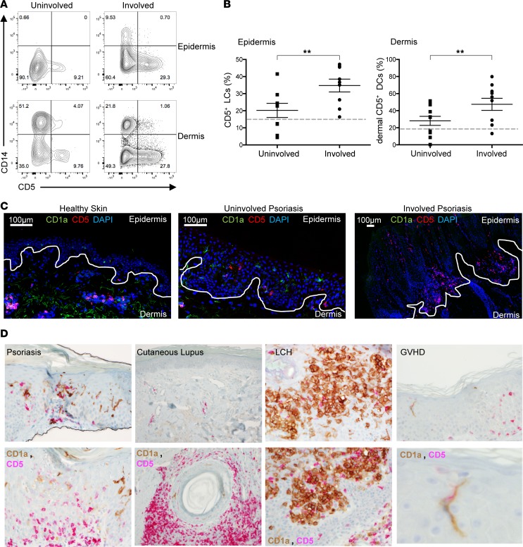
Figure 5. CD5+ LCs and dermal DCs are increased in psoriatic lesions compared with nonlesional psoriatic skin.
(A) Expression of CD5 and CD14 on epidermal and dermal DCs from uninvolved (left arm) and involved (left forearm) lesions of psoriasis patient 025 (see Table 1). (B) The percentage of CD5+ DCs in the epidermis (left) and dermis (right) of uninvolved and involved skin lesions of 7 and 8 patients, respectively. The percentage of the total migrating DCs (HLA-DR+CD3/19/56–) cells. Mean ± SEM: epidermal CD5+ DCs, uninvolved, 23.4% ± 4.6%; involved, 35.7% ± 4.7%; dermal CD5+ DCs, uninvolved, 32.7% ± 5.2%; involved, 52.2% ± 7.8%. Dashed lines mark the levels of CD5+ DCs in healthy skin. Data represent mean ± SEM; **P < 0.01 by paired Student’s t tests. (C) Immunofluorescence staining of CD5 and CD1a on healthy skin and psoriasis uninvolved and involved skin. Scale bar: 100 μM. (D) CD5 and CD1a expression in psoriasis, cutaneous lupus, Langerhans cell histiocytosis (LCH), and graft-versus-host diseased (GvHD) skin. Original magnification, ×20 (top); ×40 (bottom).

