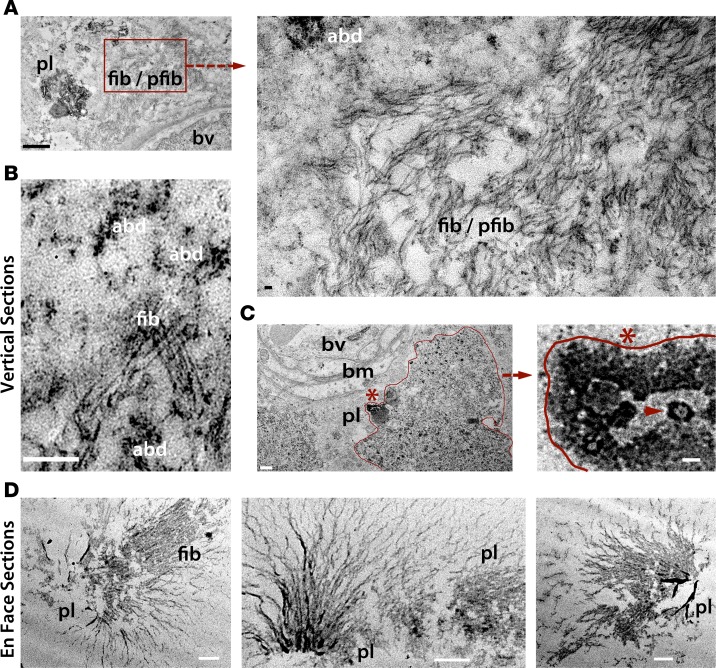
Figure 5. Ultrastructures of Aβ deposits in human AD retina.
Transmission electron microscopy (TEM) analyses of vertical (cross; A–C) and en face (horizontal; D) retinal sections from definite AD patients (n = 3; experiments repeated 3 times). Retinas were prestained with anti-Aβ42 mAb (12F4) and a high-sensitivity immunoperoxidase-based system and DAB substrate chromogen. (A) TEM image showing ultrastructure of Aβ plaque (pl), fibrils (fib), and protofibrils (pfib) near a blood vessel (bv) in vertical sections. Scale bar: 1 μm. Higher-magnification image (right), indicating presence of 10- to 150-nm-wide Aβ fibrils as well as protofibrils and Aβ deposits (abd; scale bar: 50 nm). (B) Fibrillar Aβ ultrastructure surrounded by multiple Aβ deposits. Scale bar: 200 nm. (C) Aβ plaque-like structure, marked by an asterisk and bordering red line, in close proximity to basement membrane (bm) of a blood vessel (scale bar: 0.5 μm). Higher-magnification image showing Aβ plaque-like area, with structures resembling paranuclei containing annular oligomers (arrowhead; scale bar: 40nm). (D) TEM images of en face sections demonstrating retinal Aβ plaque ultrastructures, with radial fibrillar arms emanating from a central dense core found in the innermost retinal layers. Darker black signal represents condensed Aβ aggregate core. Scale bar: 0.5 μm.

