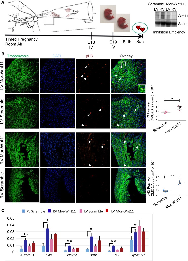
Figure 6. Systemic Wnt11 inhibition induces cardiomyocyte proliferation in a chamber-specific manner.
(A) Schematic illustration of experimental design for in vivo Wnt11 knockdown. Analysis of inhibition efficiency (Western) is shown (right panel). Mor-Wnt11 (Vivo-Morpholino-Wnt11): Wnt11-specific modified antisense oligonucleotide. (B) Representative confocal images of anti–phospho-histone H3 (anti–p-H3) immunohistochemistry (IHC) in scramble control or Mor-Wnt11–injected neonatal mouse hearts at P3. Arrows indicate representative p-H3–positive cardiomyocytes (CMCs). Original magnification, ×40. Graphs: Quantitative analysis of p-H3–positive cells (cell number/area [μm2]. (C) Expression analysis of several proliferation and mitosis markers using mRNA from LV or RV myocardium of Mor-Wnt11– or scramble-treated neonatal mouse (qRT-PCR). n = 3 per condition. Data are representative of 2 independent experiments. Error bars represent SEM. *P ≤ 0.05, **P ≤ 0.01 by unpaired, 2-tailed Student’s t test (Mor-Wnt11 compared with control).

