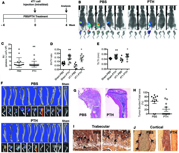
Figure 5. Intermittent PTH decreases 4T1 mouse breast cancer cell engraftment to the bone, leading to better preservation of bone architecture.
(A) Intratibial injection experimental design. Mice were 6 weeks of age at the start of the experiment and 10 weeks at the time of 4T1 cell injection. (B) Representative end point BLI images of hind limbs injected with 4T1 murine breast cancer cells. After 4 weeks, tumors were identified by BLI imaging in 7 of 15 PTH-treated mice compared with 14 of 15 in the PBS-treated mice (P = 0.005). (C) Quantitation of BLI 4 weeks after intratibial injections of 4T1 cells. Architectural analyses of proximal tibia (D) bone volume/total volume (BV/TV) and (E) trabecular thickness (Tb.Th) by μCT (n = 10). (F) Representative reconstructed 3D μCT images of tibiae with corresponding BLI images at end point in PBS- and PTH-treated mice following intratibial injections of 4T1 cells. (G) Representative H&E-stained images of tibia from PBS- and PTH-treated mice with intratibial injections of 4T1 cells (original magnification, ×4). (H) Quantitation of tumor burden. Representative TRAP staining of (I) trabecular region (original magnification, ×20) and (J) cortical region (original magnification, ×10) from tibiae of mice treated with PBS/PTH with intratibial injections of 4T1 cells. TRAP-positive osteoclasts are indicated by arrows. All values represent mean ± SD of n = 10 for each group. *P < 0.05, **P < 0.01 when compared with PBS group, by 1-way ANOVA with Bonferroni’s test as post-hoc analysis.

