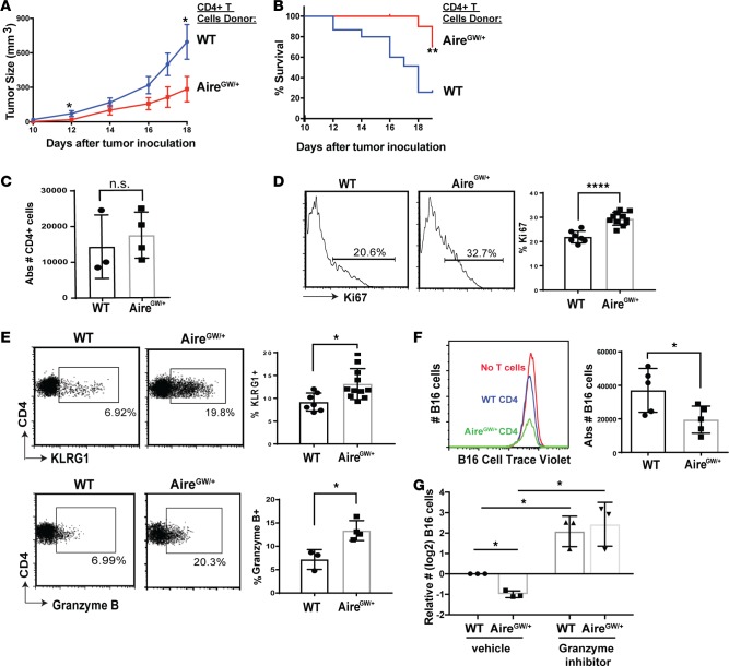
Figure 2. Aire deficiency increases CD4+ T cell cytolytic function to reduce melanoma growth.
CD4+ splenocytes were transferred from WT and AireGW/+ donors into RAG–/– recipients, followed by s.c. B16 melanoma injection on day 7. Recipients were followed for tumor growth and survival. (A and B) B16 melanoma tumor growth and survival was measured after B16 inoculation in recipients; n = 15 per group. Mann-Whitney U test. *P < 0.05, **P < 0.01. (C–E) Tumor-infiltrating lymphocytes (TIL) were harvested on day 19 following B16 melanoma inoculation in recipients of either WT or AireGW/+ CD4+ splenocytes. Absolute numbers of CD4 tumor-infiltrating cells are shown in C. Two-tailed t test. Representative flow cytometry plots and average cumulative frequencies of Ki67+ (D) and KLRG1+ and granzyme B+ (E), among CD4+ T cells. Two-tailed t test. *P < 0.05, ****P < 0.0001. (F) Representative flow cytometry histograms and average absolute CellTrace Violet–labeled B16 cell numbers after coincubation with WT and AireGW/+ CD4+ T cells. Two-tailed t test. *P < 0.05. (G) Average relative B16 cell numbers (log2) after coincubation with CD4+ T cells from WT and AireGW/+ mice, along with pan-granzyme inhibitor (3, 4 Dichloroisocoumarin). One-way ANOVA and two-tailed t tests, with P values adjusted using Hommel’s correction for multiple comparisons. *P < 0.05.

