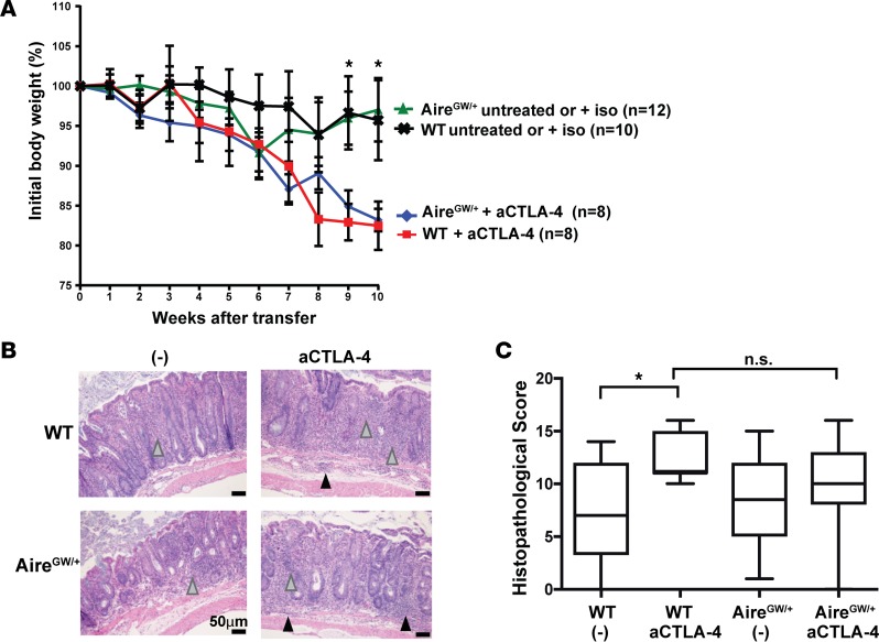
Figure 5. Aire deficiency does not exacerbate the development of experimental autoimmune colitis associated with anti–CTLA-4 antibody treatment.
(A) Percent initial body weight of RAG–/– recipients after transfer of WT or AireGW/+CD4+CD25–CD45RBhi splenocytes. After transfer, recipients were treated with anti–CTLA-4 antibody (aCTLA-4), untreated, or treated with isotype control antibody (iso). Cumulative data from 2 independent experiments are shown. *P < 0.05 comparing aCTLA-4 antibody treatment versus untreated/iso treatment in recipients of WT cells. (B) Representative H&E-stained sections of descending colons and (C) average histopathological scores of colons from recipients of CD4+CD25–CD45RBhi splenocytes derived from WT and AireGW/+ mice and administered aCTLA-4 or untreated/iso (-). Gray arrowheads, immune cell infiltration in lamina propria; black arrowheads, immune cell infiltration in the submucosa. One-way ANOVA and two-tailed t tests with P values adjusted using Hommel’s correction for multiple comparisons. *P < 0.05.

