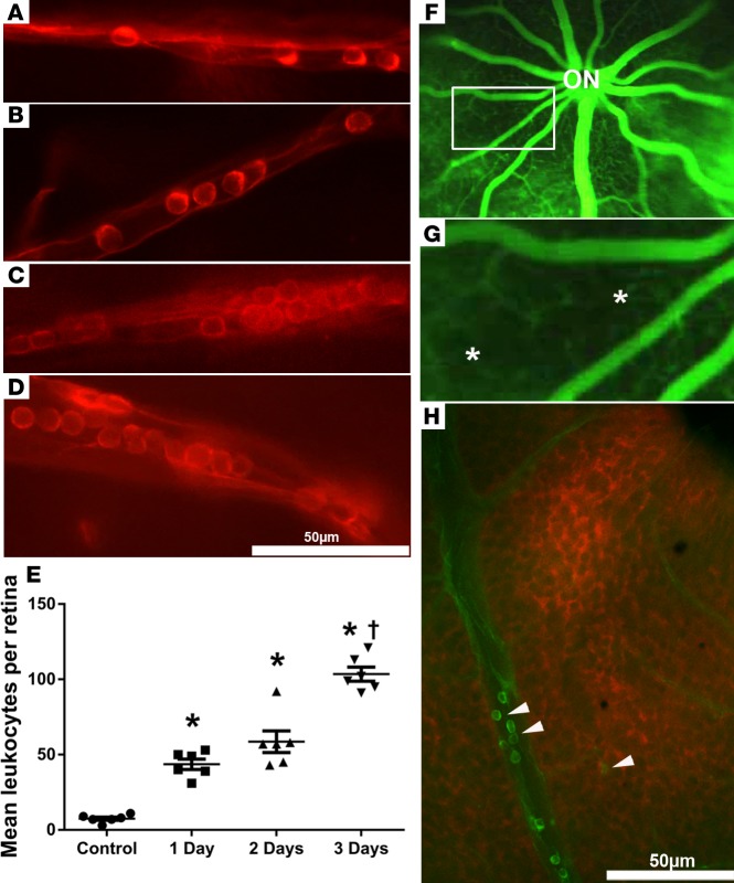
Figure 2. Sustained expression of VEGF in the retina causes leukostasis, retinal vessel closure, and retinal hypoxia.
Tet/opsin/VEGF double-transgenic mice with doxycycline-inducible expression of VEGF in the retina were given 2 mg/ml doxycycline in drinking water and perfused with rhodamine-labeled Con A 1 day (A), 2 days (B), or 3 days (C and D) after initiating doxycycline. Leukocytes were present in small vessels 1 day (A) and 2 days (B) after onset of VEGF expression and were present in vessels of all sizes after 3 days (C and D). The mean (± SEM) number of intravascular leukocytes per retina was significantly greater at each of the 3 time points compared with control (E) (*P < 0.001 by 1-way ANOVA with Bonferroni corrections) and at day 3 was greater than other time points (†P < 0.01, 1-way ANOVA with Bonferroni corrections). (F) Fluorescein angiography of Tet/opsin/VEGF mice 3 days after starting doxycycline showed dilated large retinal vessels radiating from the optic nerve (ON), between which the networks of small vessels were slightly blurred by extravascular leakage. There were hypofluorescent areas with sharp borders that appeared cut out of the diffuse fluorescence (box), better seen in a magnified view of the boxed area (G, asterisks). The dark black areas indicate absence of capillaries due to nonperfusion. Staining of a retinal flat mount with hydroxyprobe 3 days after starting doxycycline showed hypoxic retina (red) adjacent to vessels containing adherent FITC–Con A–stained leukocytes (H, arrowheads). Scale bar: 50 μm.

