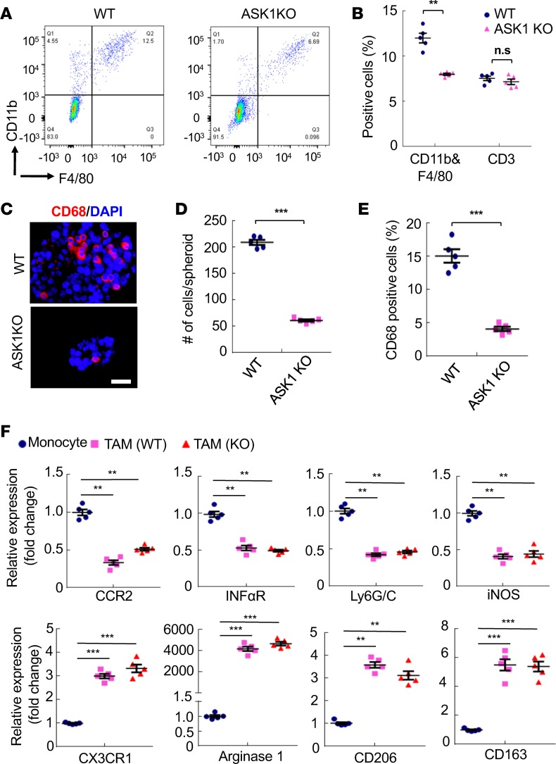
Figure 2. Attenuated peritoneal macrophage infiltration and spheroid formation in ASK1-KO mice.
In the orthotopic mouse OC model, total peritoneal cells were harvested at day 60 of WT and ASK1-KO female recipient mice. (A and B) Cells were immunostained with anti-F4/80, CD11b, and CD3e followed by FACS analyses. Isotype IgGs were used as controls. Representative images are shown in A. Percentage of F4/80+CD11b+ and CD3e+ cells in peritoneal implantations were quantified in B. Data are presented as means ± SEM, n = 5. **P < 0.01 (two-sided student’s t test). (C–E) Spheroids from ascites were collected at day 60 and mounted on slides followed by immunostaining with CD68 (C). Scale bar: 20 μm. Spheroid sizes (D) and % of CD68+ cells within spheroids (E) were quantified. (F) Accumulation of M2-like subtype tumor-associated macrophages (TAMs) had no difference between WT and ASK1-KO tumor models. F4/80+ CD11b+ macrophages in the orthotopic implantation tumor were harvested from WT and ASK1-KO mice at 8 weeks after i.p. injection of ID8 cells. M1 subtype–specific and M2 subtype–specific markers were determined by qRT-PCR. Peripheral blood monocytes were used as a control. All data are presented as means ± SEM, n = 5. **P < 0.01; ***P < 0.001 (two-sided student’s t test with no correction for multiple comparisons) compared with gene expressions in week-1 individual cells.

