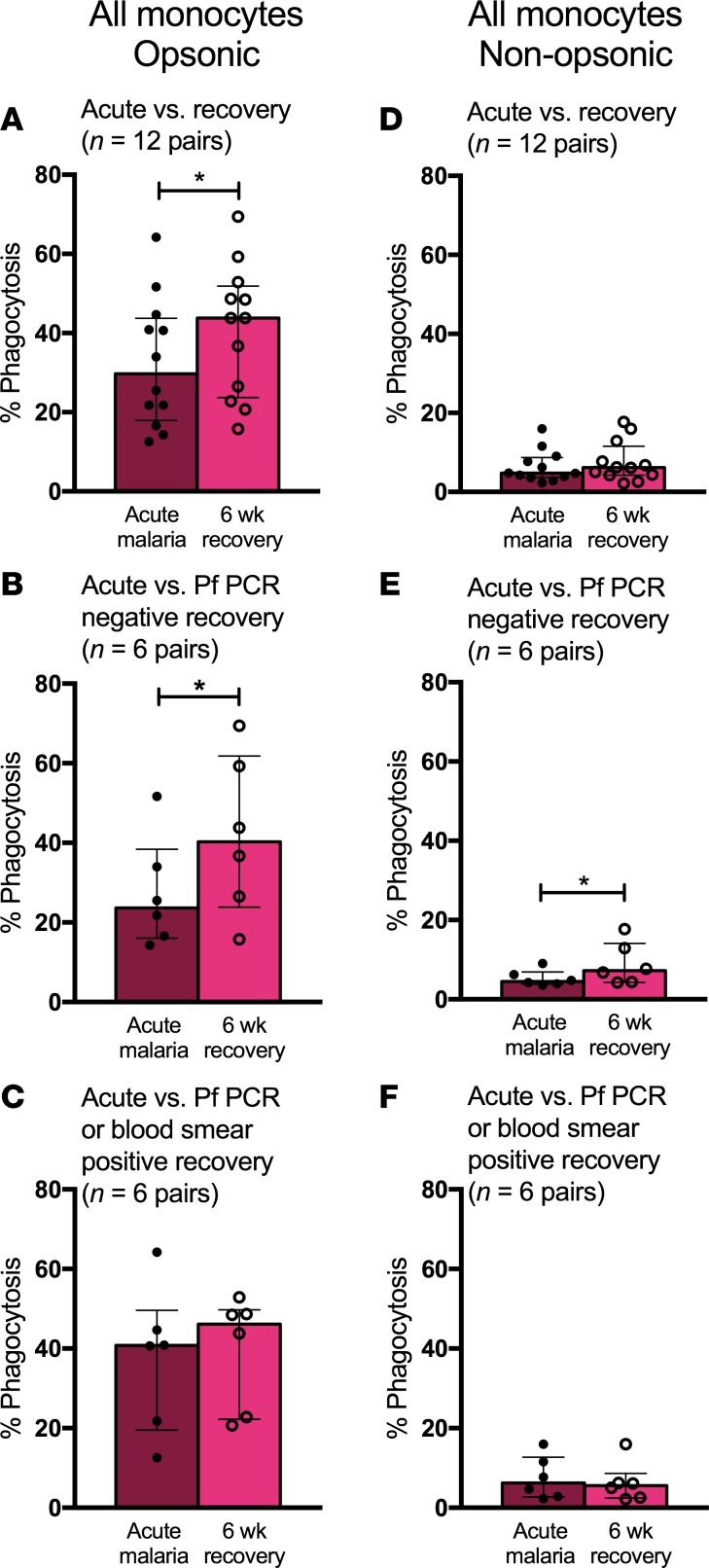
Figure 5. Monocytes display decreased phagocytic function during P. falciparum infection.
(A) Opsonic phagocytic function of all monocytes from acute malaria (AM) and 6-week recovery PBMC samples (n = 12 pairs) in the presence of Ghana14 IEs opsonized with heat-inactivated pooled plasma from Kenyan adults. (B) Opsonic phagocytic function of all monocytes from AM and 6-week PBMC samples (n = 6 pairs) from a subset of children who were P. falciparum PCR negative at 6-week follow up. (C) Opsonic phagocytic function of all monocytes from AM and 6-week PBMC samples (n = 6 pairs) from a subset of children with asymptomatic P. falciparum infection at 6-week follow up (defined by positive blood smear or P. falciparum PCR). (D) Nonopsonic phagocytic function of all monocytes from AM and 6-week PBMC samples (n = 12 pairs) in the presence of Ghana14 IEs opsonized with heat-inactivated plasma from a malaria-naive North American. (E) Nonopsonic phagocytic function of all monocytes from AM and 6-week PBMC samples (n = 6 pairs) from a subset of children who were P. falciparum PCR negative at 6-week follow up. (F) Nonopsonic phagocytic function of all monocytes from AM and 6-week PBMC samples (n = 6 pairs) from a subset of children with asymptomatic P. falciparum infection at 6-week follow up (defined by positive blood smear or P. falciparum PCR). Wilcoxon matched-pairs rank test was used to compare AM to 6-week recovery samples. Data are shown as medians with interquartile ranges. *P < 0.05.

