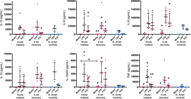
Figure 7. Monocytes from children during acute malaria and 6 weeks following treatment are highly responsive to stimulation with TLR ligands.
Monocytes were negatively selected from fresh venous blood samples from children during acute malaria and 6 weeks following treatment (n = 8 pairs). Healthy North American (N. Amer.) adult controls (n = 4) were used as experimental controls. All assays were performed with technical duplicates. Cells were cultured for 18 hours with media alone, 10 ng/ml LPS, or 100 ng/ml Pam3CSK4 (P3C), and cytokine concentrations were measured in culture supernatants. Wilcoxon matched-pairs rank test was used to compare acute malaria to 6-week recovery samples. Kruskal-Wallis test was used to compare acute malaria, 6-week recovery, and N. Amer. control samples. Data are shown as medians with interquartile ranges. *P < 0.05, **P < 0.01, as compared with controls.

