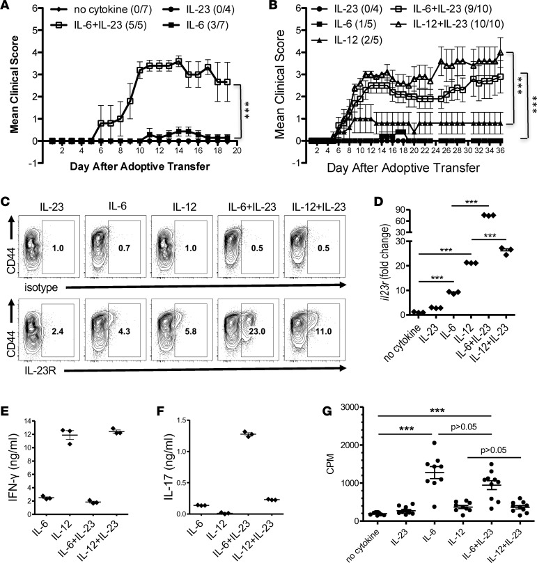
Figure 2. The combinations of IL-6+IL-23 or IL-12+IL-23 restore the encephalitogenicity to anti-CD3/CD28–activated T cells.
(A) Splenocytes from Vα2.3/Vβ8.2 TCR Tg mice were activated in vitro with anti-CD3/CD28 with or without IL-23 and/or IL-6. At 60 hours, cells were harvested and adoptively transferred into B10.PL mice (5 × 10⁶ cells/mouse). The number of mice with clinical signs/total number of mice in each group in this representative experiment is shown as follows: no cytokine (0/7); IL-23 (0/4); IL-6 (3/7); and IL-6+IL-23 (5/5). (B) Naive CD4+ T cells were purified from Vα2.3/Vβ8.2 Tg splenocytes and activated with anti-CD3/CD28 in the presence of IL-23, IL-6, and/or IL-12. At 60 hours, cells were harvested and adoptively transferred into B10.PL mice (1 × 10⁶ cells per mouse). The number of mice with clinical signs/total number of mice in each group in this representative experiment is shown as follows: IL-23 (0/4); IL-6 (1/5); IL-12 (2/5); IL-6+IL-23 (9/10); and IL-12+IL-23 (10/10). ***P < 0.001 (Mann-Whitney U test). IL-23R expression (gated on CD4+ cells) was analyzed by flow cytometry (C), and supernatants were analyzed by ELISA for IFN-γ (E) and IL-17A (F) (mean ± SEM). (D) Naive CD4+ T cells were purified from B10.PL splenocytes and activated in vitro with anti-CD3/CD28 and IL-23, IL-6, IL-12, or combinations. Cells were collected at 60 hours, and Il23r and Hprt mRNA were detected by real-time PCR. Fold change of gene expression was shown relative to no-cytokine condition (mean ± SEM). (G) Proliferation was determined by 3H-thymidine incorporation of Vα2.3/Vβ8.2 TCR Tg T cells using anti-CD3/CD28 stimulation with IL-6, IL-12, IL-23, or combinations. Each dot represents a replicate well. ***P < 0.001 (1-way ANOVA with Bonferroni’s multiple comparison test). Data is representative of ≥ 3 independent experiments.

