
Figure 4. PSGL-1–KO mice are protected from antiphospholipid antibody–mediated thrombosis.
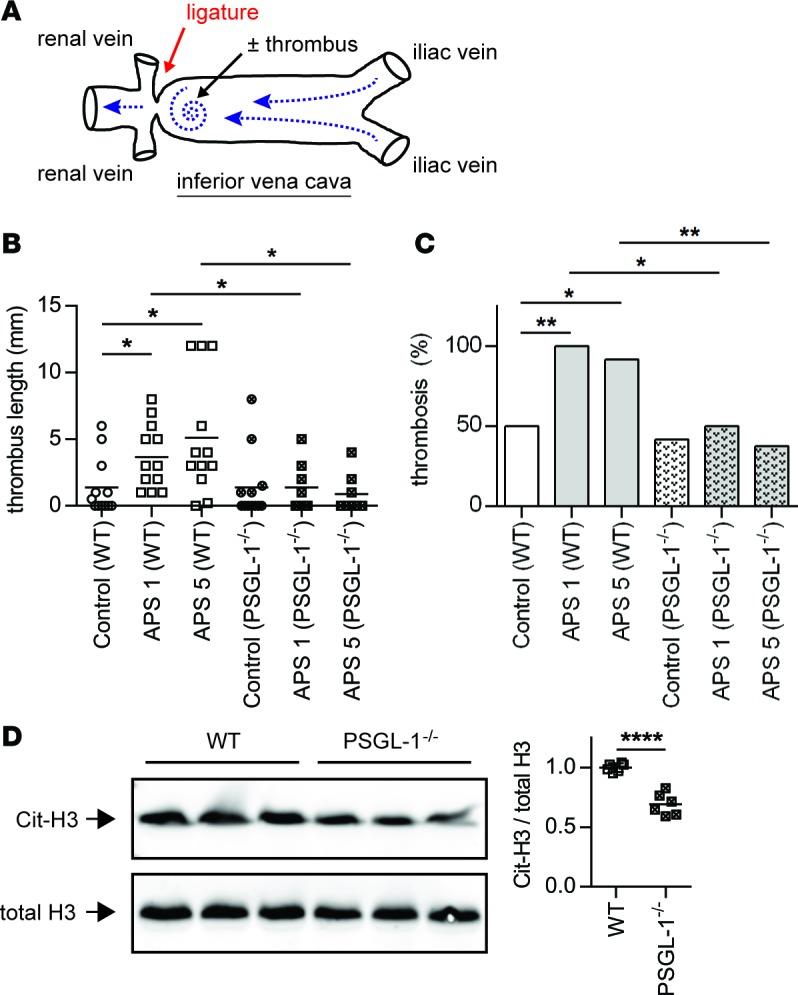
(A) Schematic of flow-restriction model of venous thrombosis. Mice were treated with IgG isolated from either healthy controls or patients with primary APS as described in Methods. The inferior vena cava (IVC) was then narrowed by fixing a ligature around the IVC (using a spacer to ensure uniformity). When a thrombus formed, it was just distal to the stenosis, in the area indicated by the spiral. (B) Thrombus length was measured 6 hours after flow restriction in WT or PSGL-1–/– mice. APS 1 and APS 5 denote IgG from two different patients. The mean for each group is denoted by a horizontal line, while each data point represents an individual mouse; *P < 0.05 adjusted using Kruskal-Wallis test followed by Dunn’s multiple comparisons test. (C) This is a contingency analysis of the data presented in panel B. *P < 0.05 and **P < 0.01 by χ2 test. (D) NET content of APS thrombi (presented in panel B) was assessed by Western blotting for citrullinated histone H3 (Cit-H3) and total histone H3 (the two blots were run in parallel/contemporaneously). Quantification is based on n = 6 per group (3 each from APS 1 and APS 5); ****P < 0.0001 by two-tailed t test.