
Figure 5. Antiphospholipid antibodies promote increased leukocyte adhesion to the inferior vena cava following flow restriction.
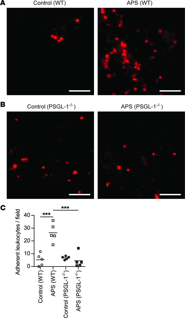
Mice were treated with control or APS IgG (specifically APS 1 as in Figure 4). (A) WT mice. Representative images of the inferior vena cava, 1 hour after flow restriction. Leukocytes are stained red by Rhodamine 6G. Scale bars: 50 μm. (B) PSGL-1–/– mice. Representative images and quantification of adherent leukocytes as described in panel A. Scale bars: 50 μm. (C) Quantification of adherent leukocytes per 40× field. The mean for each group is denoted by a horizontal line, while each data point represents a unique mouse; ***P < 0.001 adjusted using one-way ANOVA followed by Tukey’s multiple comparisons test.