
Figure 1. Kinase domain architecture and dynamics.
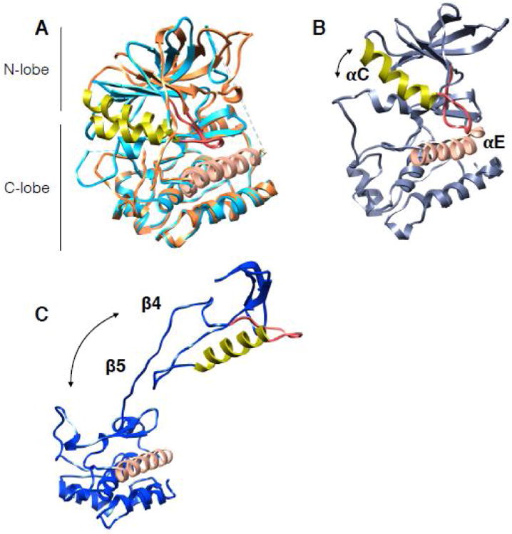
The αC helix in all structures is in lime color, adjacent αC-β4 loop is in red. The proteins have been aligned by the αE helix (salmon color helix in C-lobe). Arrows show the direction and relative magnitude of N-lobe motion in relation to C-lobe. (A) Structures of EGFR in the active state (light blue in color, PDB:2ITP) and the inactive state (piper in color, PDB:2GS7). Note the small shift in αC helix between states. (B) Snapshot from 12us all-atom MD simulation on EGFR from Shaw group displaying an opening between the kinase lobes. (C) Cdk4 kinase structure as it is in the cryoEM complex of Hsp90-Cdc37-Cdk4 (PDB:5FWL) showing dramatic unfolding between the kinase lobes and unraveling of β4- β5 strands.