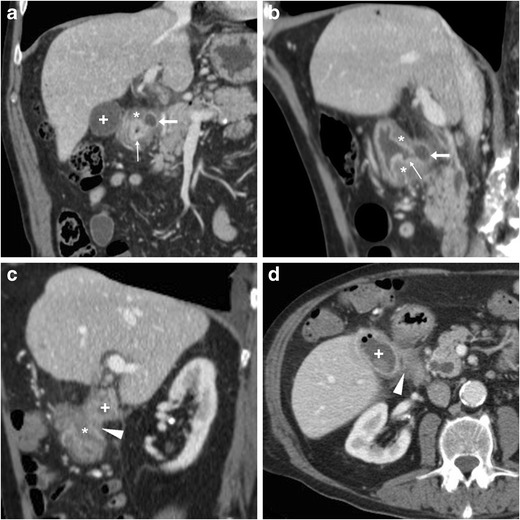
Fig. 18.

A 78-year-old male with history of chronic NSAID use had initial contrast-enhanced CT (a, b) finding of circumferential thickening of pylorus and proximal duodenum with oedematous submucosa (*), enhancing mucosa (thin arrows) and posterior ulcer outpouching (arrows); ventrally, the normal-appearing gallbladder (+) was in contact with the affected duodenal bulb. Fifteen months later, the patient experienced melaena and abdominal pain: repeated CT (c, d) showed development of communication (arrowheads) between thickened duodenum (*) and contracted gallbladder (+) with intraluminal air and some dependent sludge or blood
