Figure 1.
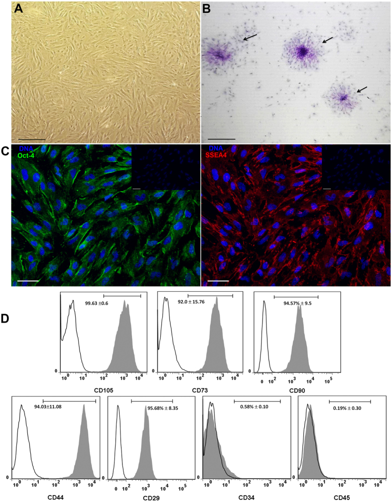
Cells obtained from human amniotic membrane mesoderm displayed mesenchymal stromal cells characteristics. Phase-contrast micrograph of hAMSC adhered to a polystyrene cell culture plate at 3rd passage showing fibroblast morphology; the photograph was taken at 40x of magnification, scale bar 100 μm (A). The cells were cultured for ten days and stained with crystal violet, and a direct light micrograph was performed in order to identify the UFC (arrows); the photograph was magnified at 35x in a stereoscopic microscope, scale bar 100 μm (B). Fluorescence micrographs of hAMSC stained with pluripotent embryonic markers OCT-4 (left panel), and SSEA-4 (right panel). DAPI was used to identify their nuclei in both panels; scale bars represent 20 μm (C). hAMSC cells from 4th passage were trypsinized and stained with antibodies against the indicated cell surface antigens and analyzed by flow cytometry. As shown, cells were positive to (>90%) CD105, CD73, CD90, CD44 and CD29; in contrast, they were negative to the expression of CD34 and CD45 hematopoietic-cells markers, inner numbers represent the mean ± SD (D). These are representative images from three different independent assays.

