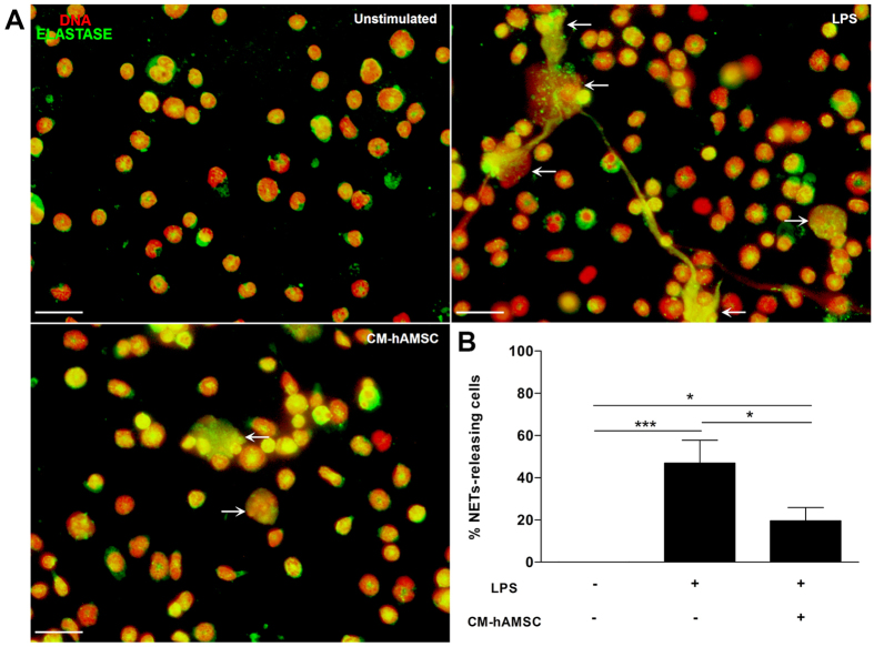
Figure 3.
The soluble factors from hAMSC decrease the release of NETs. Murine neutrophils isolated from bone marrow were stimulated with LPS to induce the release of NETs and were incubated with CM from hAMSC. Fluorescence micrographs of unstimulated neutrophils (upper-left panel), LPS-stimulated neutrophils (upper-right panel, LPS) and LPS-neutrophils cultured with the CM from hAMSC (lower-left panel, CM-hAMSC). LPS-stimulated neutrophils liberated extracellular traps formed by elastase and DNA (white arrows). The neutrophils in contact with the soluble factors from hAMSC (CM-hAMSC) decrease the liberation of NETs. Scale bar represents 20 μm. These are representative images from three independent assays (A). Graphic represents the percentage of NETs releasing cells. The area of NETs was quantified with the Image J program from five random fields in each condition. Bars represent the mean percentage of NETs releasing cells ± SD (n = 3), *p < 0.05 (LPS vs. CM-hAMSC; unstimulated vs. CM-hAMSC); ***p < 0.001 (unstimulated vs. LPS) (B).

