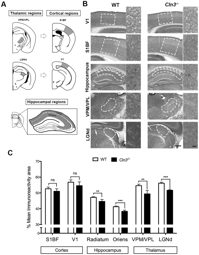
Figure 1.
Spatio-temporal synaptic loss study in Cln3 −/− detected differentially vulnerable synaptic populations across brain regions. (A) Brain region schematic showing the brain areas measured in grey. Thalamic regions includes the ventral posterior medial/ventral posterior lateral thalamic nucleus (VPM/VPL) (top left) and the dorsal lateral geniculate nucleus (LGNd) (bottom left); their respective cortical projections in the primary somatosensory cortex (S1BF) (top right) and primary visual cortex (V1) respectively (bottom right); hippocampal regions measured within the CA1-3 were the stratum radiatum and stratum oriens (bottom). (B and C) Representative photomicrographs of coronal sections of the same brain regions immunostained with synaptophysin (Syp) and bar chart showing its corresponding quantification based on the area of immunoreactivity in 13 month old control and Cln3 −/− mice. Syp immunoreactivity was lower in thalamic regions (VPM/VPL and LGNd) in the Cln3 −/− mice when compared to controls indicating more pathology, detectable earlier in the thalamus. Hippocampal stratum oriens and stratum radiatum also showed reduced Syp immunostaining, although the difference between genotypes was smaller. Cortical regions did not show difference in immunoreactivity for synaptophysin indicating that no synaptic loss is happening in these cortical areas at 13 months. (Mean ± SEM; *P < 0.05; **P < 0.01; ***P < 0.001; ns P > 0.05, Student T test, 5 mice per each genotype and time-point were used, Scale bar = 200 um (left) and 20 um (right).

