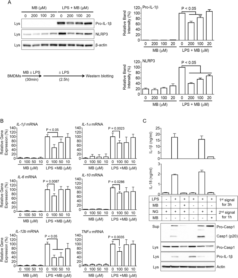
Figure 2.
Effect of MB on the priming step of inflammasome activation and expression of other cytokine genes. (A and B) BMDMs were treated with the indicated concentration of MB with/without LPS (10 ng/mL) as indicated in the schematic graph. (A) Pro-IL-1β and NLRP3 expression levels were analyzed by immunoblotting and further presented with band density. (B) Expression levels of mouse IL-1β, IL-1α, IL-6, IL-10, IL-12b, and TNFα mRNAs were quantitated by real-time PCR. C, BMDMs were treated with MB and/or LPS as the 1st signal, after which cells were replaced by media containing nigericin (NG, 2nd signal) with/without MB as the 2nd signal. IL-1β and IL-18 secretion levels were measured by ELISA, and Casp1 secretion and pro-IL1β expression were analyzed by immunoblotting. All immunoblot data shown are representative of at least three independent experiments. Bar graph presents the mean ± SD.

