Figure 4.
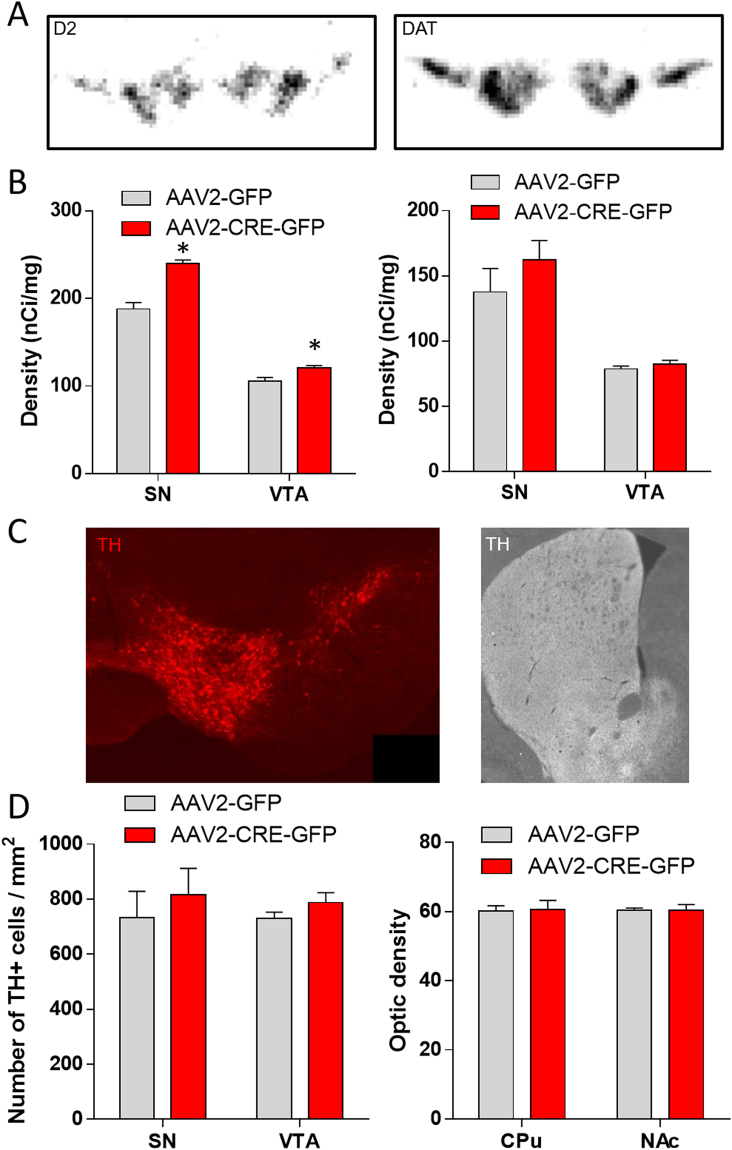
Neuroanatomical consequences of nigrostriatal DA depletion. (A) Illustration of D2 (left) and DAT (right) mRNA radioactive in situ labelling in the SN and the VTA. (B) Density (nCi/mg) of D2 (left) and DAT (right) mRNA expression in the SN and VTA, measured by radioactive in situ hybridization, 16-week after AAV2-GFP or AAV2-CRE-GFP bilateral injection in VMAT2lox/lox mice (n = 4 per groups, Mann-Whitney U: AAV2-GFP vs AAV2-CRE-GFP: D2: SN *p < 0.05; VTA *p < 0.05; DAT: SN p = 0.47; VTA p = 0.14). (C) Illustration of TH immunostaining in DA neurons of the SN and the VTA (Left) and in the fibers in the CPu and NAc (Right). (D) Number of TH positive cell per mm2 in the SN and the VTA (Left) and optic density of TH positive fibers in the CPu and the NAc (Right) 16-week after AAV2-GFP or AAV2-CRE-GFP bilateral injection in VMAT2lox/lox mice (n = 4 per groups, Mann-Whitney U: AAV2-GFP vs AAV2-CRE-GFP: TH + cells: SN p = 0.59; VTA p = 0.38; optic density: CPu p = 0.86; NAc p = 0.6). SN: Substancia nigra, VTA: Ventral Tegmental Area, CPu: Caudate Putamen, NAc: Nucleus Accumbens, PFC: PreFrontal Cortex, VMAT2: Vesicular Monoamine Transporter-2, AAV: Adenoassociated Virus, TH: Tyrosine hydroxylase.

