Figure 1.
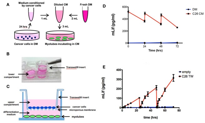
Cell culture models of tumor cell treated myotubes and the associated LIF levels in medium. (A) Schematic drawing of the design for the conventional conditioned medium (CM) model. C26 cancer cells were incubated in differentiation medium (DM) for 24 h to produce conditioned medium. Four day-differentiated C2C12 myotubes were treated for 72 h with 33% CM in fresh DM, refreshed every 24 h. (B) Photograph of a Transwell insert being placed into a well of a 6 well plate. (C) Schematic diagram of the Transwell system with C26 cancer cells in the upper compartment and C2C12 4-day myotubes growing below (figure is redrawn from Corning website). Cancer cells were seeded on microporous membrane insert (0.4 μm) placed into the well containing myotubes for 72 h with the upper and lower compartments containing DM, refreshed every 24 h. (D) Graph of LIF levels in medium from myotube cultures treated with DM (control) or C26 CM at the beginning and end of each 24 h period of treatment. (E) Graph of LIF levels in the lower compartment of myotube cultures with Transwell (TW) inserts seeded with C26 cells. “Empty” indicates control myotube cultures containing Transwell inserts without C26 cells.

