
Figure 5.
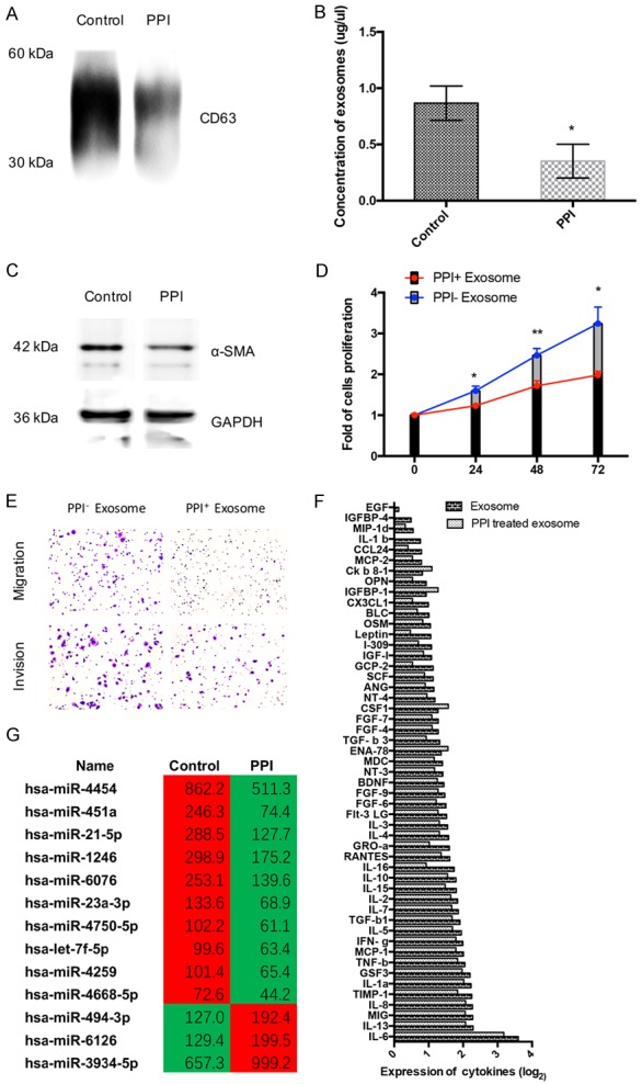
PPIs inhibited the release of exosomes and exosomes related miRNA and regulated tumor microenvironment though regulating exosomes. A. PPIs (80 ug/ml, 24 h) effect on the expression CD63 in the exosome of SGC7901 cells (n=3). B. PPI (80 ug/ml, 24 h) effect on the concentration of exosomes were measured by BCA protein assay (n=5). C. HFF-1 cell were treated with exosome of SGC7901 cells (80 ug/ml of PPI or PBS (control) treated for 24 h), and the expression of α-SMA and β-actin of HFF-1 cells were tested by Western Bloting (n=3). D. SGC7901 cells were cultured with the medium of HFF-1 cell, which were treated with exosome of SGC7901 cells (80 ug/ml of PPI or PBS (control) treated for 24 h). CCK8 assay was performed to analyze cell proliferation of SGC-7901 cells (n=5). E. SGC7901 cells were cultured with the medium of HFF-1 cell, which were treated with exosome of SGC7901 cells (80 ug/ml of PPI or PBS (control) treated for 24 h). Migration and invasion of SGC-7901 cells were tested by transwell assays (n=3). F. HFF-1 cell were treated with exosome of SGC7901 cells (80 ug/ml of PPI or PBS (control) treated for 24 h), and secreted cytokine were tested by Human Cytokine Array (n=3). G. The unsupervised hierarchical clustering analysis (microarray) of de-regulated miRNAs of exosome. The left is control group and right is the PPIs group. (|Fold change| ≥ 1.5 and P < 0.05) (n=3). All photographs were taken at a magnification of ×200. Asterisk indicates a significant difference determined by unpaired two-tailed t test (*** indicates P < 0.001; ** indicates P < 0.01; * indicates P < 0.05).