
Figure 1.
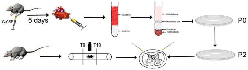
Process of isolation, culture transplantation of PBMSCs. SD rats weighing 80-120 g were subcutaneously injected with G-CSF for 6 d. About 6 ml of peripheral blood was harvested from the left ventricle after anesthetization. Then the sample was diluted with a same volume of PBS and immediately overlaid on rat Ficoll lymphocyte separation liquid. After centrifugation, the cells were cultured until at 80%-90% confluence and then subcultured. Adult rats underwent a contusion of spinal cord for model control after dorsal laminectomy. Following dorsal laminectomy at T9-T10 level, spinal cord was contused with a new type spinal cord impactor. Thirty minutes after SCI, 10 μl of PBS and PBMSC suspension (2×10⁴ cells/μl) were respectively injected into each rat in both the PBS and PBMSCT groups at 2 μl/min and the injection sites were adjacent rostral and caudal spinal cord at a distance of 0.05 mm from the lesion.