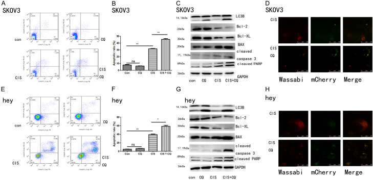
Figure 4.
The combination of chloroquine and cisplatin on cell apoptosis and autophagy in SKOV3 and hey cells. Apoptosis was evaluated with PE Annexin V and 7-ADD staining in SKOV3 and hey cells after treatment with cisplatin (5 uM for both SKOV3 and hey cells) and/or chloroquine (10 uM for SKOV3 cells and 5 uM for hey cells) for 24 hours. (A and E) Representative dot plots illustrating the data near the mean of the groups in (B and F). *, P < 0.05 and **, P < 0.01 for comparisons between groups. (C and G) SKOV3 and hey cells were treated with cisplatin (5 uM for both SKOV3 and hey cells) and/or chloroquine (10 uM for SKOV3 cells and 5 uM for hey cells) for 24 hours. Western blotting analysis of LC3B, Bcl-2, Bcl-XL, Bax, cleaved caspase 3 and cleaved PARP. Western blotting of GAPDH was included as a loading control. (D and H) SKOV3 and hey cells stably expressing mCherry-Wassabi-LC3B were treated with cisplatin (5 uM for both SKOV3 and hey cells) and/or chloroquine (10 uM for SKOV3 cells and 5 uM for hey cells) for 24 hours, then subjected to confocal microscope analysis. Green-positive, red-positive puncta (G+R+) are autophagosomes; green-negative, red positive puncta (G-R+) are autophagolysosomes.

