
Figure 5.
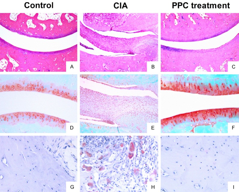
The pathological manifestations of CIA rats after PPC treatment. All animals were sacrificed at day 42 after primary immunization. Ankle joints were harvested for evaluating the pathological changes. A-C: Showed the representative results of HE staining. A: The articular surface was smooth without any infiltration of inflammatory cells in the articular cavity of control group. B: The infiltration of inflammatory cells and the unlimited proliferation of SF cells in the articular cavity of CIA rats. C: The articular surface was smooth without invading SF cells after PPC treatment. D-F: Showed the results of Safranin O-fast green staining. D: The bone structure and cartilage of control group were intact in articular cavity. E: The articular cartilage encountered serious destruction in CIA rats. F: The condition of destruction was ameliorated by PPC treatment. G-I: Showed the results of TRAP staining. G: Only a few osteoclasts were found in control joints. H: The numbers of osteoclasts were significantly increased in the joints of CIA rats. I: The numbers of osteoclasts were obviously decreased after PPC treatment.