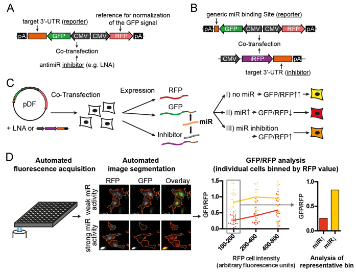
Figure 1.
Schematic representation of dual-fluorescent reporter assays. (A and B) Reporter constructs for the determination of miRNA activity in cells are cotransfected with two types of inhibitors: the effect of an miRNA on a target is assessed by cloning the UTR of interest downstream of GFP and treatment with small molecule inhibitors (LNA-antimiRs, A). Conversely, the inhibitory effect of a UTR on an miRNA can be assessed by cloning generic binding sites downstream of GFP and co-transfecting plasmids expressing the UTR of interest downstream of the non-interfering fluorophore iRFP (infrared fluorescent protein (36), B). (C) MiRNA activity is assessed by measuring the GFP normalized to the RFP (tdTomato) signal after reporter transfection into cells. (D) Readouts for GFP and RFP values for individual cells are obtained by automated microscopic image acquisition and high content image analysis. Selection of cells from a narrow range with little miRNA inhibition by the reporter reduces variability due to different transfection strength between cells.

