Fig. 5.

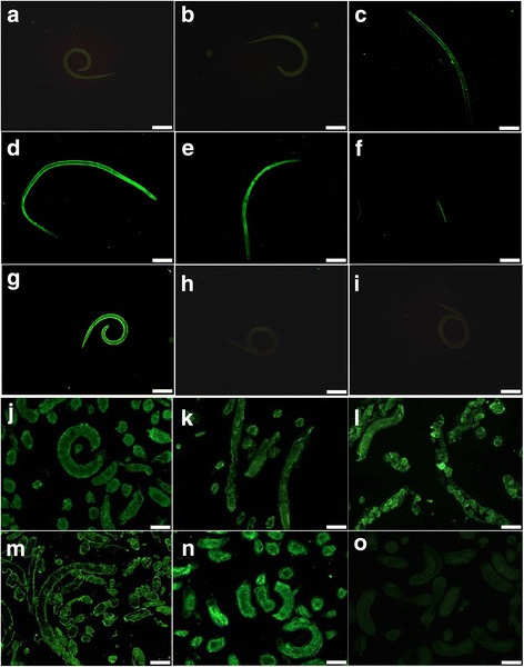
Expression and immunolocalization of TspGST at Trichinella spiralis various stages. a-i The results of IIFT with the intact parasites probed by anti-rTspGST sera. The obvious fluorescent staining is observed on the surface of AW at 3 (c) and 6 dpi (d, e), and NBL (f), but not on the surface of ML (a) and IIL (b). The ML recognized by sera from T. spiralis experimentally infected mice g was used as a positive control; the ML incubated with normal mouse sera (h), and PBS (i) were used as negative controls. j-o Sections of intact worms (ML, IIL and AW) reacted with anti-rTspGST sera. The immunostaining is observed at the cuticle of ML (j), IIL (k), AW at 3 (l) and 6 dpi (m), especially at embryos within the female uterus. The ML recognized by sera from T. spiralis experimentally infected mice n as positive control; the ML incubated with normal mouse sera o as negative control. Scale-bars: a-e, g-i, 200 μm; f, j-o, 100 μm
