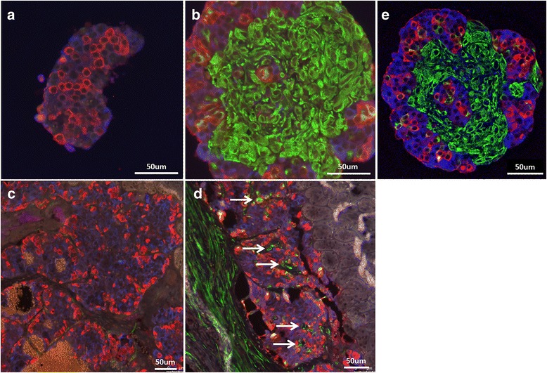
Fig. 2.

Alpha and beta cell arrangement in cultured pseudoislets and after transplantation in SCID mice. Islets were dissociated by enzymatic digestion, and 104 single cells were cultured in hanging drops for 6 days prior to in vitro immunofluorescence studies, and for 8 days prior to transplantation under the kidney capsule of SCID mice. Beta cells were immunostained for insulin (blue), alpha cells for glucagon (red), and MSC for vimentin (green). Representative images of three different experiments. a, b Pseudoislets after in vitro formation: (a) islet cells alone, (b) islet cells and MSC. c, d Pseudoislets 15 days after transplantation: (c) pseudoislets composed of islet cells alone, (d) pseudoislets composed of islet cells and MSC. White arrows indicate vimentin-positive cells within the islet graft located outside of the alpha and beta cell arrangements (islet substructures). e Right panel: in vitro formed islets and MSC at day 6, confocal laser scanning microscopy
