Fig. 3.

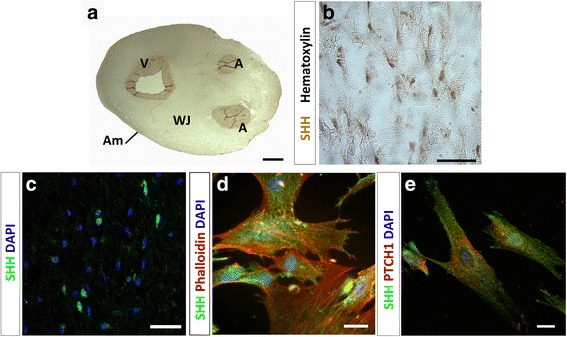
SHH is expressed in human umbilical cord. a Representative image of immunohistochemistry against SHH in an umbilical cord section. b Imuunohistochemistry shows positive immunoreactivity in cells immersed in the WJ, with hematoxylin staining allowing cell nuclei visualization. c SHH+ cells are detected in the WJ by immunofluorescence. d In primary cultures of WJ-MSC, SHH was detected at the cellular surface. e Double immunostaining reveals coexpression of SHH and PTCH1 in WJ-MSC suggesting an autocrine signaling. Scale bars = 0.5 cm (a), 50 μm (b, c), and 20 μm (d, e). A artery, Am amnios, DAPI 4’,6-diamidino-2-phenylindole, PTCH1 Patched1, SHH Sonic hedgehog, V vein, WJ Wharton’s jelly
