
Fig. 4.
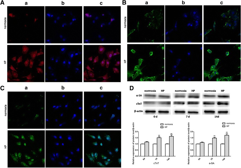
Cardiogenic differentiation of CSCs. (A), (B), (C) Confocal microscopy of immunofluorescent staining of DAPI-labeled CSCs induced by 5-AZA after 14 days (400×). (A) Immunofluorescent staining of apelin: (a) cells stained with antibody to apelin appeared red; (b) cells derived from DAPI-labeled CSCs induced by 5-AZA displayed blue nuclei; (c) merged image of a and b. (B), (C) Immunofluorescent staining of α-SA and cTnT respectively: (a) cells stained with antibody to α-SA and cTnT appeared green; (b) cells derived from DAPI-labeled CSCs induced by 5-AZA displayed blue nuclei; (c) merged image of a and b. (D) Alterations of the protein expressions of cardiogenic differentiation specific genes. Western blot analysis to detect alterations of protein expressions of cardiogenic differentiation specific genes including α-SA and cTnT at different time points after induction of cardiogenic differentiation. Comparisons were made between normoxia and HP groups. **p < 0.01 vs normoxia. HP: hypoxia preconditioning