Fig. 3.

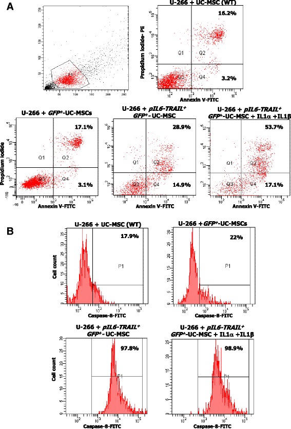
In vitro apoptosis of U-266 cells induced by pIL6-TRAIL + -GFP +-UC-MSCs. a Apoptosis in U-266 cells by transduced UC-MSCs was measured by Annexin V/PI staining using flow cytometry. Representative dot plots revealed that the apoptosis extent was significantly increased (43.8%) after 24 h of coculture with pIL6-TRAIL + -GFP +-UC-MSCs with respect to control UC-MSCs (19.4%) and GFP +-UC-MSCs (20.2%). The effect was also enhanced when IL-1α and IL-1β were added to the cocultures (70.9% of U-266 cell apoptosis). b Active caspase-8 in U-266 cells as signature of TRAIL-induced apoptosis was measured by flow cytometry after 24 h of coculture with UC-MSCs, GFP +-UC-MSCs, and pIL6-TRAIL + -GFP +-UC-MSCs. This representative experiment depicts the activity of caspase-8 in 97.8% of U266 cells cocultured with pIL6-TRAIL + -GFP +-UC-MSCs as compared to 17.9% of control UC-MSCs, and 22% of GFP +-UC-MSCs. Such high levels of active caspase-8 were not further modified by supplementing the cultures with IL-1α/IL-1β (98.9% of positive cells). GFP green fluorescent protein, MSC mesenchymal/stromal stem cell, pIL6 interleukin-6 promoter, TRAIL tumor necrosis factor related apoptosis inducing ligand, UC umbilical cord, WT wildtype
