
Fig. 2.
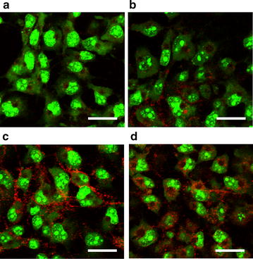
PfCSP gene expression following transduction of the BDES vaccine into mammalian cells. a–d COS-7 cells were transduced with AcNPV-WT (a), BDES-PfCSP1-gp64 (b), BDES-sPfCSP2-Spier (c) and BDES-sPfCSP2-Spider (d) each at a multiplicity of infection of 500. After 48 h incubation, the cells were stained with the 2A10 mAb conjugated to Alexa Fluor 594 (red). Counterstaining of the live cells were conducted by the SYTO-13 nucleic acid dye (green). Bars, 50 µm