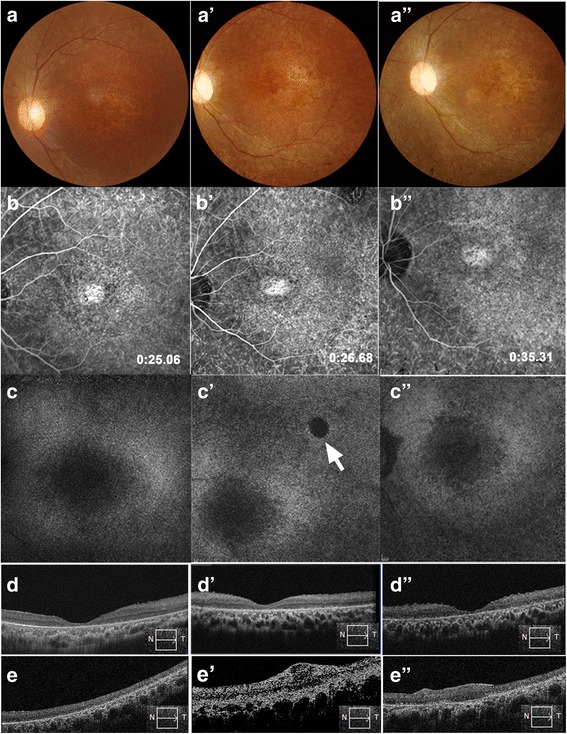
Fig. 4.

Retinal morphological changes after RPC transplantation in patient 6. a–d Baseline images, a’–d’ 12-month follow-up, and a”–d” 24-month follow-up. a Color fundus photographs, b fluorescein angiograms, c autofluorescence imaging in the macular area, d foveal optical coherence tomography (OCT), and e horizontal OCT scanning along the injection site. a’,a” No retinal hemorrhage or edema occurred after RPC transplantation. b’,b” The characteristics of fluorescent leakage did not change after transplantation. c’,c” No obvious autofluorescence destruction in the macular area after RPC transplantation, except for a minimal area of hypo-autofluorescence (arrow). This disrupted RPE layer corresponded to the injection site. d’,d” Foveal depression was maintained pre- and postoperatively, indicating that no macular edema occurred. e The injection site before surgery (box and arrow indicate the direction of OCT scanning). e’,e” Signs of the injected cells could not be observed at 12 and 24 months post-implant and, in this patient, retinal scarring was evident with local retinal thickening
