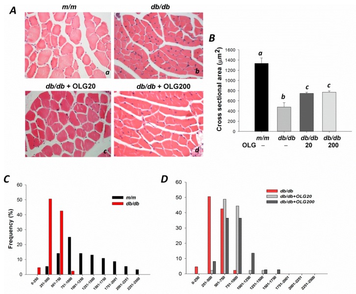
Figure 1.
The cross-sectional area of tibialis anterior muscle in m/m, db/db mice, and db/db mice with oligonol (OLG) supplementation. (Aa–d) Myofibers were stained with hematoxylin-eosin (400×); (B) Mean cross-sectional area of tibialis anterior muscle; (C) The distribution of myofiber sizes in tibialis anterior muscles from m/m and db/db mice; and (D) db/db mice with or without oligonol (OLG) supplement.

