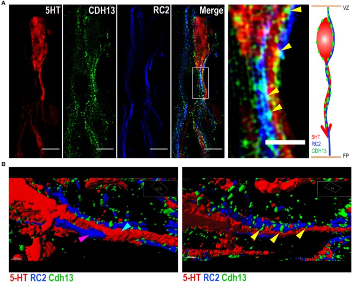
Figure 7.
Cdh13 at points of intersection between 5-HT neurons and radial glial cells. Representative images of serotonergic fibers and radial glial cell (RGC) extensions triple-stained for 5-HT, RC2 and Cdh13. (A,B) Cdh13 (green) is present in both 5-HT neurons (red) and RGCs (blue), and at some points of intersection between these two cell types (yellow arrows). (B) Reconstruction of triple IF of 5-HT, RC2, and Cdh13 using Imaris. The 5-HT neuron (cyan arrow) is intertwined with the RGC fiber (magenta arrow). Cdh13 immunoreactivity is found at the interface between both cell types (Supplemental Videos 1 and 2). Orientation: sagittal. Scale bars in (A) 5 μm in full images, 2 μm in the magnified boxed region. FP, floor plate; VZ, ventricular zone.

