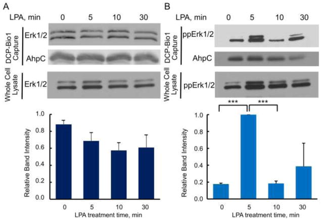
Figure 5.
ERK1/2 oxidation in ovarian cancer-derived SK-OV-3 cells treated with lysophosphatidic acid (LPA). SK-OV-3 cells depleted of serum for 18 h were treated with 100 nM LPA and harvested in the presence of DCP-Bio1 as described in Figures 1 and 2. The upper panels show representative immunoblots of DCP-Bio1 labeled proteins for total ERK (A) and TEY-phosphorylated ERK1/2 (B). The lower panels depict averaged relative intensity for each sample after LPA treatment, normalized as in Figure 4 (n=6 for total ERK, n=3 for phospho-ERK). *** p<0.001

