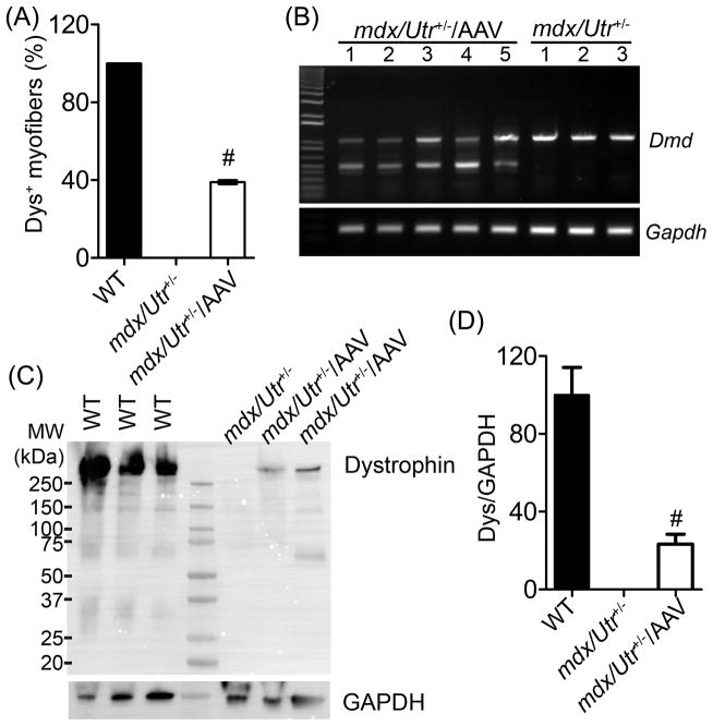
Figure 3. Restoration of dystrophin in the cardiomyocytes of mdx/Utr+/− mice.
(A) Measurement of dystrophin-positive cardiomyocytes (% of total caveolin-3-positive cardiomyocytes; mean ± SD) in mdx/Utr+/− heart cryosections treated with or without 1 × 10¹² vg AAV-SaCas9/gRNAs (N=4 per group). (B) RT-PCR analysis of heart tissues from mdx/Utr+/− mice treated with (N=5) or without (N=3) 1 × 10¹² vg AAV-SaCas9/gRNA. Genome editing is expected to produce a smaller (~500 bp) dystrophin band. (C) Immunoblotting of heart lysates from WT, mdx/Utr+/− and mdx/Utr+/− with AAV-SaCas9/gRNAs using anti-dystrophin and anti-GAPDH antibodies. (D) Quantitative analysis of dystrophin expression by immunoblotting of heart lysates from wild-type (WT), mdx/Utr+/− and AAV treated mdx/Utr+/− (N=3 per group). # p < 0.01.

